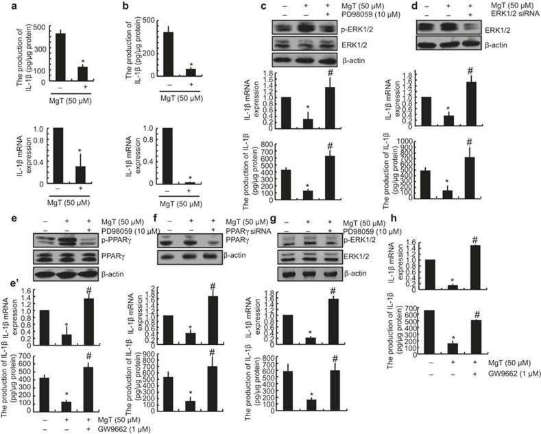
Figure 3.
Involvement of ERK1/2 and PPARγ pathways in regulating the expression of IL-1β in MgT-treated A172 or D1A cells. Human glioblastoma A172 (a) or mouse astrocytes/microglia D1A cells (b) were treated with MgT (50 μM) for 48 h. In select experiments, A172 cells were treated with PD98059 (10 μM) in the absence or presence of MgT (50 μM) for 48 h (c and e). In separate experiments, the cells were transfected with ERK1/2 (d) or PPARγ siRNA (f) before incubation with MgT (50 μM) for 48 h. In distinct experiments, A172 cells were treated with GW9662 (1 μM) in the absence or presence of MgT (50 μM) for 48 h (e'). In other experiments, primary cultured astrocytes were treated with MgT (50 μM) in the absence or presence of PD98059 (10 μM) (g) or GW9662 (1 μM) for 48 h (h). Total ERK1/2 (c, d and g upper panel), phosphorylated ERK1/2 levels (c and g upper panel), total PPARγ (e and f upper panel), and phosphorylated PPARγ (e) were detected by immunoblotting using specific Abs. Equal lane loading is demonstrated by the similar intensities of total β-actin. IL-1β protein and mRNA levels were determined by IL-1β enzyme immunoassay kits and qRT-PCR, respectively. The total amounts of protein and GAPDH served as internal controls. The data represent the means ± SE of three independent experiments. *P < 0.05 compared with the vehicle-treated control. #P < 0.05 compared with MgT treatment alone.

