Figure 7.
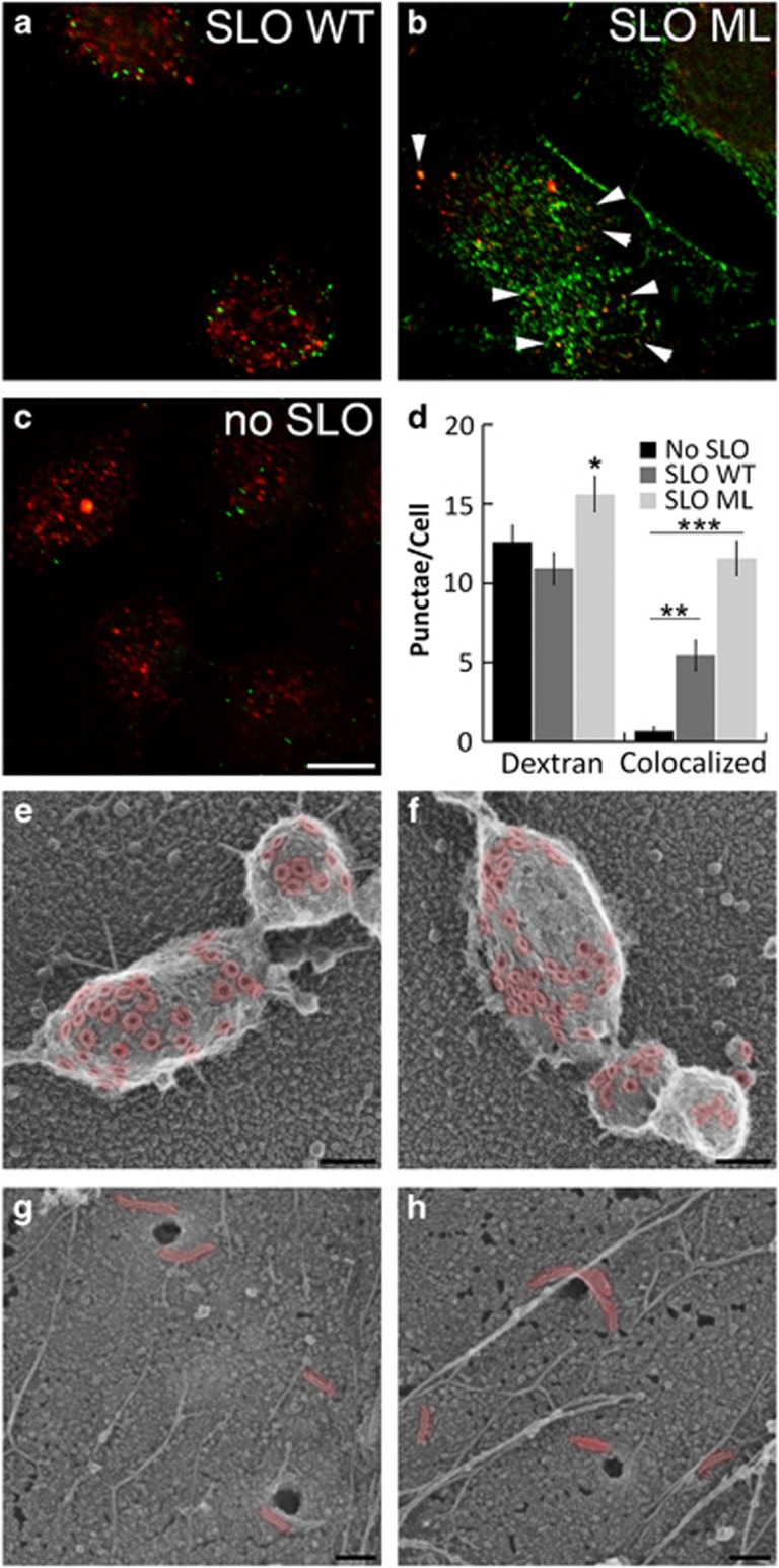
Compensatory endocytosis removes inactive SLO from the cell surface. (a–d) HeLa cells were seeded onto coverslips, serum starved for 1 h in RPMI and challenged with sublytic dose of SLO WT (a), the equivalent mass of SLO ML (b), or no toxin (c) in the presence of 2.5 mg/ml dextran Texas Red (red) in RC for 15 min at 37 °C. Coverslips were fixed, permeabilized, and stained with 6D11 anti-SLO antibody followed by anti-mouse 488 antibody, mounted and analyzed by confocal microscopy. (d) The number of dextran punctae and dextran/SLO double-positive punctae was quantified by direct counts. (e–h) HeLa cells were seeded onto coverslips, challenged with a sublytic dose of SLO (e and f) or mass equivalent of SLO ML (g and h) for 5 min at 37 °C, washed, fixed in 2% glutaraldehyde and analyzed by rapid-freeze, ‘deep-etch' EM. SLO pores and oligomers are pseudo-colored red to aid visualization. Immunofluorescent images show one representative image of five independent experiments. The graph displays the average±S.E.M. of five independent experiments. EM micrographs show representative fields from two independent experiments. *P<0.05, **P<0.01 and ***P<0.001. Scale bar=10 μm for (a–c) and 100 nm for (e–h)

