Figure 1.

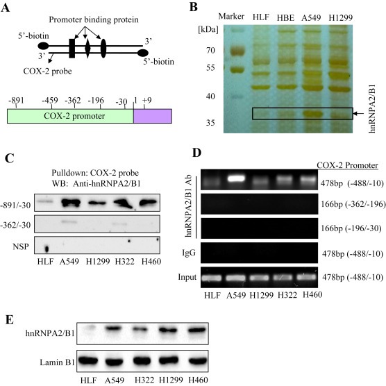
Binding of hnRNPA2/B1 to the COX‐2 promoter. (A) The COX‐2 promoter probe and structure. (B) SDS‐PAGE and silver staining of COX‐2 promoter binding proteins. (C) Binding of hnRNPA2/B1 to the biotinylated COX‐2 promoter probe. The anti‐hnRNPA2/B1 antibody was used to validate hnRNPA2/B1 protein in COX‐2 probe‐streptavidin bead complex by Western blot. NSP, a non‐specific DNA probe. (D) Binding of hnRNPA2/B1 to the COX‐2 promoter in chromatin structure by ChIP assay. The complex of hnRNPA2/B1 antibody‐chromatin were immunoprecipitated, and the PCR products were amplified using the different primers of COX‐2 promoter. IgG, a negative control for ChIP assay. (E) Detection of hnRNPA2/B1 expression in cell nuclear extracts. LaminB1, a loading control for nuclear proteins.
