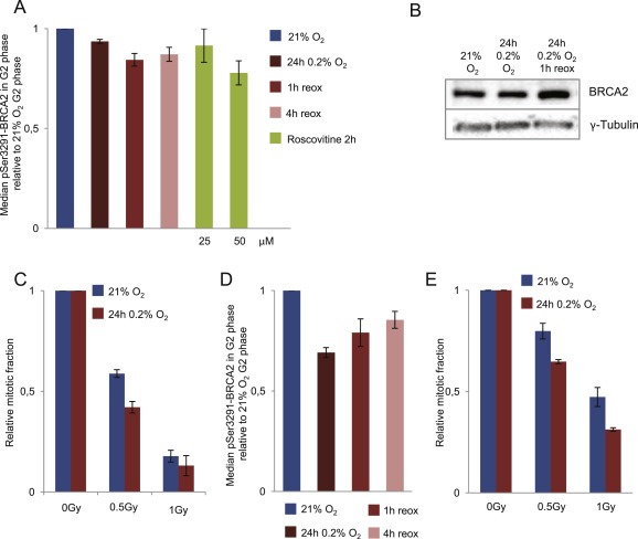
Figure 3.

Hypoxia‐induced changes in CDK activity and G2 checkpoint activation. A. CDK activity in G2 phase cells as measured by phosphorylation of BRCA2‐Ser3291. U2OS cells were grown at 21% O2, or incubated at 0.2% O2 for 24 h and harvested inside the hypoxia chamber and at 1 and 4 h after reoxygenation, or treated with Roscovitine for 2 h at 21% O2. Flow cytometry barcoding analysis of phospho‐BRCA2‐Ser3291 was performed as in Figure 2 and S2. B. Immunoblot analysis of U2OS cells treated as in A with antibodies to total BRCA2 and γ‐tubulin (loading control). C. G2 checkpoint activation after IR (0.2%O2 24 h). Flow cytometric analysis of G2 checkpoint arrest after X‐ray irradiation (0, 0.5, 1 Gy) of normoxic U2OS cells (21%O2) or U2OS cells exposed to 24 h of hypoxia at 0.2%O2 and irradiated 15 min after reoxygenation. Nocodazole was added to all samples 1 h after IR, and the samples were harvested 5 h later. The relative mitotic fraction was determined as the fraction of phospho‐H3 positive cells in irradiated samples divided by the fraction of phospho‐H3 positive cells in non‐irradiated samples. Average values from 3 independent experiments are shown. Error bars indicate SEM. D. Phosphorylation of BRCA2‐Ser3291 in H460 cells treated with hypoxia and analyzed as in A. E. G2 checkpoint activation in H460 cells treated with hypoxia and IR and analyzed as in C.
