
Figure 3.
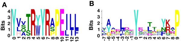
Motif analysis of hyperphosphorylated tyrosine sites. A) A motif identified by MEME for hyperphosphorylated tyrosine residues. The zero position for the motif indicates the hyperphosphorylated tyrosine residue. B) A motif identified by GLAM2 for hyperphosphorylated tyrosine residues. The zero position for the motif indicates the hyperphosphorylated tyrosine residue.