Figure 2.

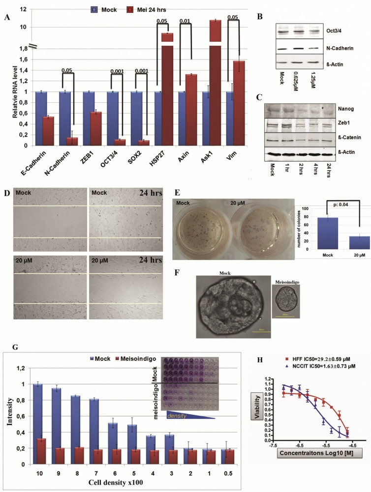
Meisoindigo repressed CSC phenotypes in Jopaca‐1. (A) Meisoindigo suppressed CSC‐associated gene expression analyzed by qRT‐PCR in Jopaca‐1 cells at 20 μM for 24 h. (B) Meisoindigo repressed CSC‐associated proteins. Jopaca‐1 cells were incubated with meisoindigo (0.625 μM and 1.25 μM) for 48 h. The whole cell lysate was subjected to immunoblot. Specific antibodies against Oct3/4 and N‐Cadherin were used. β‐Actin served as loading control and DMSO as mock. (C) Meisoindigo inhibited activities of CSC‐associated proteins in a time‐dependant manner. Jopaca‐1 cells were incubated with meisoindigo (20 μM) as indicated and analyzed by immunoblot. (D) Meisoindigo inhibited gap closure in wound healing assay of Jopaca‐1 cells treated at 20 μM concentration. Photos were taken at the beginning and after 24 h treatment. (E) Meisoindigo inhibited colony formation in soft agar assay. Jopaca‐1 cells were pre‐treated with meisoindigo (20 μM) or DMSO for 24 h. After removal of compound the cells were re‐plated in agar‐coated plates. The number of colonies (>50 cells) was scored using microscopy images. (F) Meisoindigo reduced cell self‐renewal in mammosphere assay (scale bar: 50 μm). (G) Meisoindigo reduced cellular clonogenic potential in the in vitro dilution assay. After 24 h treatment with 20 μM meisoindigo, cells were re‐plated in 96‐well plate at indicated cell density. Colonies were allowed to form within 14 days without changing the medium thereafter stained with MTT and counted by microscopy. The number of colonies was normalized to that obtained from plating 1000 cells. The dye was dissolved in DMSO and wells were photographed as depicted in the figure. (H) Cell viabilities of HFF and NCCIT were evaluated in a 72 h MTT assay.
