Figure 3.

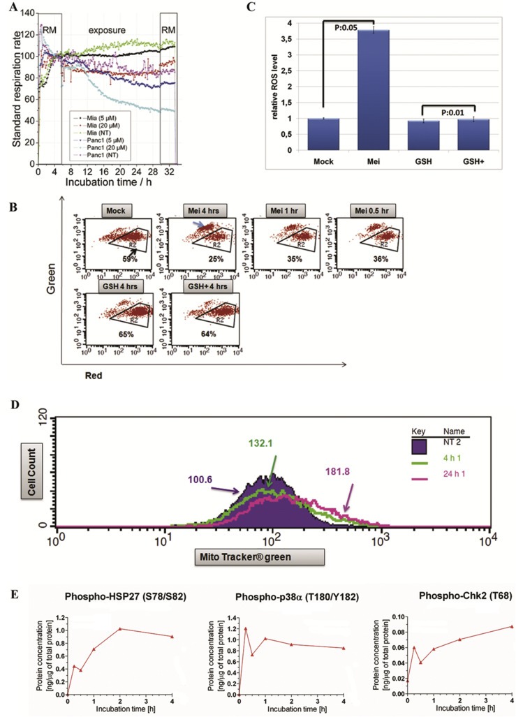
Meisoindigo regulated mitochondrial activity. (A) Meisoindigo reduced cell respiration in Miapaca2 and Panc1 cells monitored by Bionas 2500. The cells were incubated in Bionas running medium (RM) for 6 h and treated with compound at two different concentrations for 24 h. The data of cellular respiration was normalized to corresponding mock treatment. (B) Meisoindigo reduced mitochondrial membrane potential. Jopaca‐1 cells were incubated with meisoindigo (20 μM), antioxidant glutathione (GSH, 5 mM) or with a combination (all combination of agents with meisoindigo were abbreviated as agent+). The cells were collected at indicated time points, stained with JC‐1 and analyzed by FACS. Region R2 was gated to indicate redhigh/greenlow signal, while blue arrow pointed to redlow/greenhigh signal. (C) Meisoindigo enhanced cellular ROS formation. The level of ROS was determined by using ROS specific dye DHE in Jopaca‐1 treated with DMSO as mock or meisoindigo 20 μM for 4 h. (D) Total amount of mitochondrial mass was increased upon meisoindigo. Mito tracker green was used to stain mitochondria in living cells incubated with meisoindigo 20 μM for 4 h and 24 h and analyzed by FACS. The mean value of mitochondrial mass is highlighted. (E) Meisoindigo activated p38, HSP27 and CHK2 analyzed by protein microarray.
