
Figure 4.
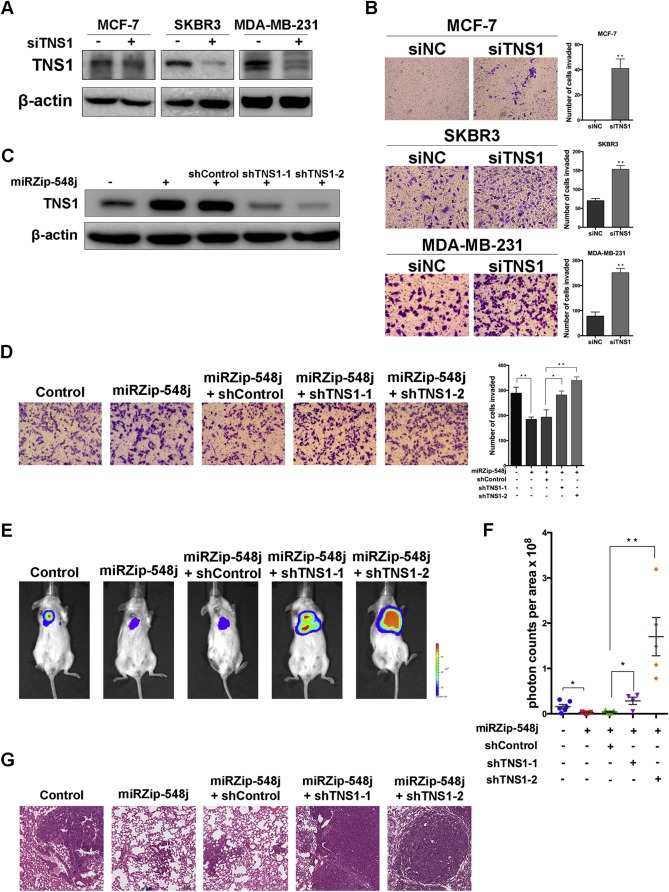
Tensin1 is involved in the regulation of cell invasion and metastasis by miR‐548j. (A) Tensin1 protein levels in MCF‐7, SKBR3 and MDA‐MB‐231 cells transfected with control siRNA (siNC) or Tensin1 siRNA (siTNS1) were analyzed by Western blotting. β‐actin is shown as a loading control. (B) Transwell invasion assays were performed to examine the invasiveness of the indicated cells after transfection with control siRNA (siNC) or Tensin1 siRNA (siTNS1). Results are quantified (right panel). (C) Western blot of Tensin1 protein levels in 231‐Luc cells infected with a control or miRZip‐548j together with a control shRNA or one of two different shRNAs targeting Tensin1. (D) Representative micrographs and quantification of ‘Rescue’ experiments performed by transwell invasion assays. (E) Representative photos of bioluminescence signals of mice from each group in ‘Rescue’ experiments. In vivo metastasis was assayed as described in Materials and Methods. (F) Quantitative analysis of metastasis. The values of the bioluminescence signals from each group were quantified and analyzed using the Mann–Whitney U‐test. (G) Lung metastasis in mice in which the indicated cells were implanted was confirmed by H&E staining. Error bars in B, D and F represent mean ± SEM. *P < 0.05, **P < 0.01.