Figure 4.

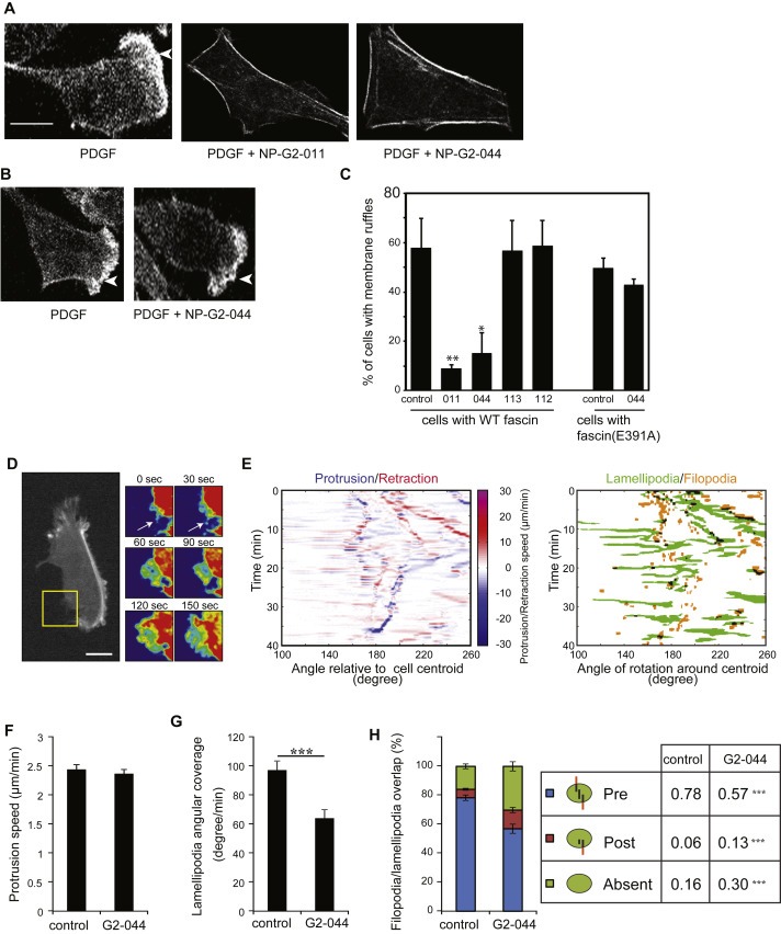
Inhibitory effect of Compounds NP‐G2‐011 and NP‐G2‐044 on lamellipodial formation and the transition from filopodia to lamellipodia. (A) Cells with wild‐type fascin were treated with PDGF to induce lamellipodial formation (marked by arrowheads). Addition of Compound NP‐G2‐011 or NP‐G2‐044 blocked PDGF‐induced lamellipodial formation. (B) 4T1 cells expressing fascin(E391A) mutant formed lamellipodia upon PDGF treatment. Lamellipodial formation in these cells was resistant to Compound NP‐G2‐044 inhibition. (C) Quantification of data in A and B. Error bars, mean ± SEM. *p < 0.05; **p < 0.01. Scale bar, 10 μm. (D) Time lapse of a LifeAct‐GFP expressing MDA‐MB‐231 cell showing a filopodium (arrow at 0 and 30 s) extending as a lamellipodium (60–150 s in zoom of yellow box sown at right). (E) Angular projection map of the protrusion and retraction speed of cell membrane extensions along with the identified lamellipodial and filopodial structure map. (F) Average protruding speed of membrane extension with and without NP‐G2‐044 treatment. (G) Quantification of the angular membrane fraction covered by lamellipodia with and without NP‐G2‐044 treatment. (H) Quantification of the lamellipodial and filopodial overlap with and without NP‐G2‐044 treatment. (Pre, presence of a filopodium prior to lamellipodial extension; Post, overlap of a filopodium after the extension of a lamellipodium; Absent, no overlap between filopodia and lamellipodia). Error bars, mean ± s.e.m. Scale bars, 10 μm ***p < 0.01.
