
Figure 6.
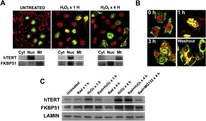
Oxidative stress promotes hTERT nuclear export to mitochondria. (A) HeLa cells were transfected with pCI‐Neo‐hEST2‐HA, treated with 0.5 mM H2O2 for 1 h or 4 h, and the subcellular localization of endogenous FKBP51 (red) and HA‐hTERT (green) was visualized by confocal microscopy. Western blots show the localization of both proteins after a subcellular fractionation into cytosol (Cyt), nuclei (Nuc) and mitochondria (Mt). (B) HeLa cells were treated with 0.5 mM H2O2 for 1 h, 3 h, or 3 h followed by an extra 1 h incubation without peroxide (4 h total). The reversion of the subcellular redistribution of endogenous hTERT (red) and endogenous FKBP51 (green) was evaluated by confocal microscopy. (C) Cells were treated as described in the figure and 50 μg proteins were resolved by Western blot. Lamin B was used as loading control.