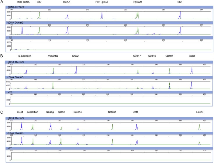
Figure 1.

Multiplex‐RT‐PCR for the detection of epithelial, EMT and stem cell markers. Depicted are electropherograms of three multiplex‐RT‐PCR panels after analysis by capillary electrophoresis. For all panels, upper row: gDNA, middle row: cDNA, lower row: ‐RT. Primer specificity and PCR‐performance was successfully validated on genomic DNA and cDNA of OvCar3 cells. A) Epithelial marker panel: CK7 (124 bp), Muc‐1 (149 bp), EpCAM (222 bp) and CK5. PDH (100/183 bp) was used as control to distinguish between gDNA and cDNA. B) EMT marker panel: N‐cadherin (127 bp), Vimentin (170 bp), Snai2 (208 bp), CD117 (309 bp), CD146 (335 bp), CD49f (363 bp) and Snai1 (402 bp). C) Stem cell marker panel: CD44 (120 bp), ALDH1A1 (139 bp), Nanog (162 bp), SOX2 (185 bp), Notch4 (210 bp), Notch1 (268 bp), Oct4 (310 bp) and Lin28 (413 bp).
