
Figure 5.
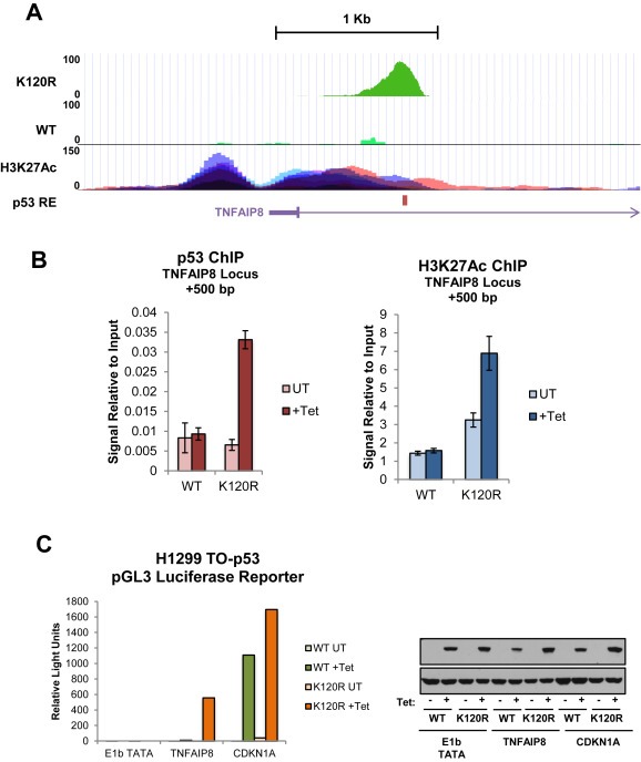
TNFAIP8 locus contains a functional, cryptic p53 response element (A) Higher resolution of TNFAIP8 locus shown in Figure 4A with ChIP‐Seq peaks for WT and K120R (Tetracycline treated condition only), location of p53 response element (RE) is indicated by red bar. H3K27Ac marks on seven cell lines (peaks are distinguished by color) from ENCODE are also shown (Encode Project Consortium, 2012). (B) ChIP signal at TNFAIP8 locus (+500 bp), for p53 or H3K27Ac from DNA extracted from cells as described in Figure 4. Signal is normalized to input DNA. (C) A luciferase reporter plasmid containing either the p53 response element associated with CDKN1A or TNFAIP8 was transfected into TO‐p53 WT or K120R cells. The plasmid containing the E1b TATA only was included as a negative control. After 24 h of tetracycline treatment, cells were harvested and luminescence was measured using the Nano‐Glo Luciferase Assay System. Right panel: Western blot showing p53 induction for cells used in reporter assay.