
Figure 1. Expression of plasminogen activators and plasminogen at the site of pulmonary emboli.
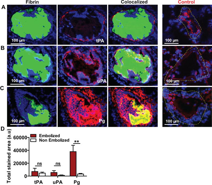
Lung tissue containing FITC-fibrin labeled (green) pulmonary emboli was immunostained (red) to detect the expression of (A) tPA, (B) uPA and (C) plasminogen (Pg) (D) The total immune-stained area (arbitrary units; a.u) for each protein was measured in a 20× (100 μm) image of an embolized vs non embolized (Control; dashed yellow outline) pulmonary artery in the lungs. n=3, mean ± SEM. **p<0.01; ns, non-significant.