
Figure 4.
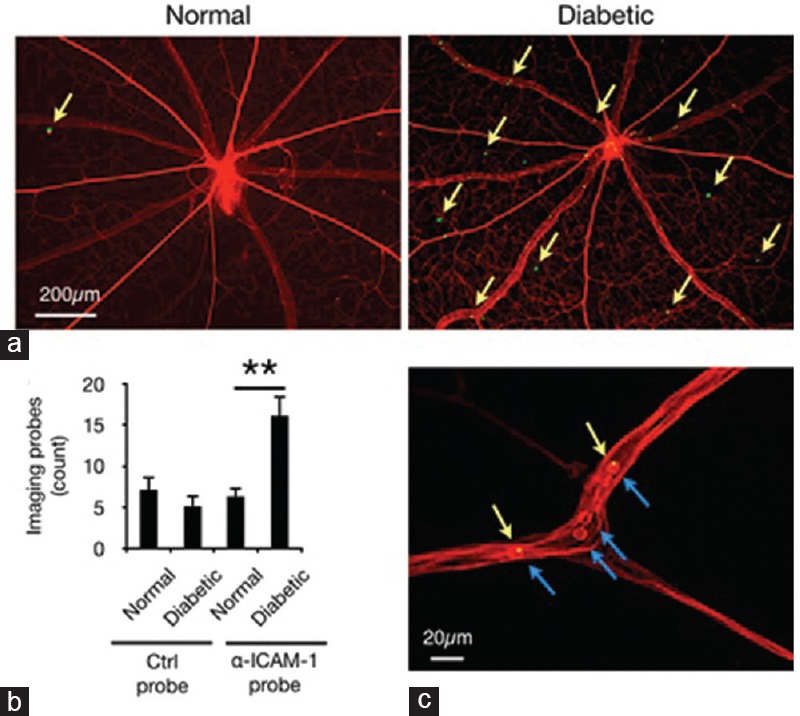
Ex-vivo evaluation of imaging probe accumulation. Retinal flatmounts were prepared from animals perfused with rhodamine-ConA (red). Imaging probes that resisted perfusion are visible as green spots in fluorescence microscopy. (a) Representative retinal micrographs show probe adhesion in retinal vessels of normal and diabetic rats. (b) Quantification of α-ICAM-1 probe adhesion in retinal vessels (n = 6–8, **P < 0.01). (c) Imaging probes bound to firmly adhering leukocytes in retinal vessels. Micrograph shows rhodamine-stained leukocytes (blue arrows) and a-ICAM-1 probes (green/yellow).