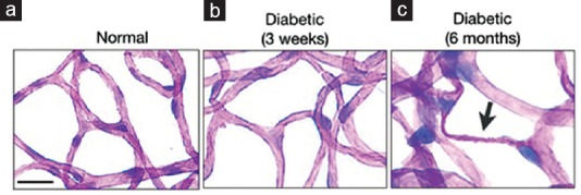
Figure 5.

Early detection by molecular imaging precedes structural damage. Retinas of normal and diabetic animals were trypsin-digested to visualize vascular changes in the diabetic retina. PAS and hematoxylin-stained flatmounts of trypsin-digested normal retinas show patent retinal capillaries, which are comprised of endothelial cells and are surrounded by pericytes (a). At three weeks of diabetes, retinas of diabetic animals show no signs of structural damage (b). In contrast, long-term diabetic animals (six months) display obliterated acellular capillaries (arrow) (c). Bar, 50 μm.
