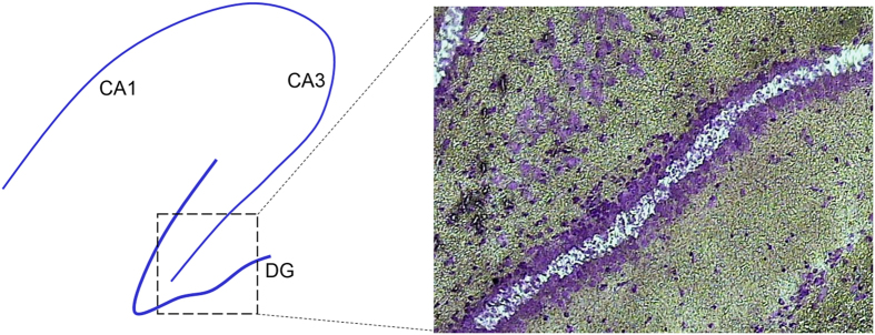
Figure 2. Laser capture harvesting of cells in the middle of the dentate granule cell layer in the outer leaf.
Left: diagram of hippocampus with CA1 and CA3 pyramidal cell layers and the dentate gyrus (DG). Laser capture was done from the boxed region and shown in the right panel. Cresyl violet stain of a section from a rat treated with pilocarpine 10 days before. The region of captured dentate granule cells is indicated by the absence of blue stain.

