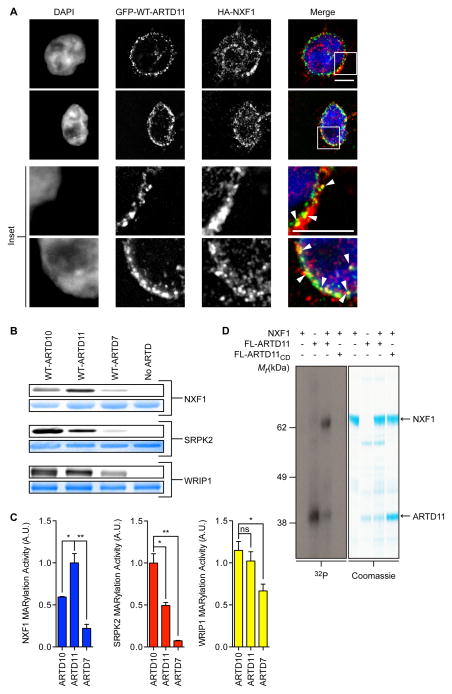
Figure 5. Validation of MARylation Targets Identified via GFP-IG-ARTD – Modified 6-a-NAD+ Pairs.
(A) GFP-ARTD11 and HA-NXF1 are partially co-localized at the nuclear membrane. HEK 293T cells co-expressing GFP-ARTD11 and HA-NXF1 were fixed with paraformaldehyde and processed for immunofluorescence. DNA was stained with DAPI. Scale bar = 5 μm. Inset: white arrowheads show co-localization.
(B) In vitro WT-ARTDcat MARylation assays demonstrate that NXF1 is a preferred ARTD11 substrate. WT-ARTD10cat, -ARTD11cat, and -ARTD7cat were screened for MARylation activity using recombinant NXF1, SRPK2, and WRIP1 in the presence of 6-a-NAD+. The same gel was first fluorescently imaged to detect substrate MARylation (top gel, gray) and then stained to detect total substrate (bottom gel, blue).
(C) Quantification of results shown in (B). The bar graphs below depict the MARylation activity for each substrate with each WT-ARTD (mean ± S.E.M., n=3). (*) represents p-value < 0.05 and (**) represents p-value < 0.01, two-tailed student t-test. ns = not significant.
(D) Results from NXF1 in vitro MARylation assay using full-length ARTD11 and 32P-NAD+. Full-length ARTD11 or ARTD11CD was incubated with NXF1 in the presence of 32P-NAD+ and ADPr transfer was visualized using autoradiography (left, gray) and stained to detect total substrate (right, blue). Arrows indicate FL-ARTD11 and NXF1.
See also Figures S4 and S5.

