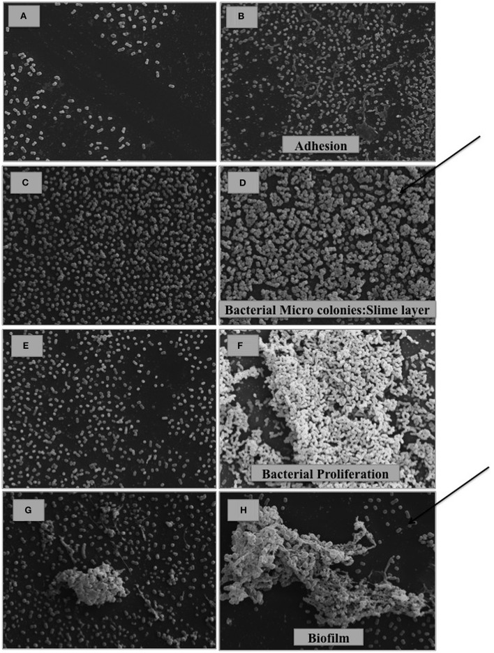
Figure 2.
SEM analysis of A. baumannii cells cultured in the absence (A,C,E,G) and presence of 0.5% bile salts (B,D,F,H). (A,B) A. baumannii ATCC 17978; (C,D) A. baumannii ΔadeB ATCC 17978; (E,F) A. baumannii ΔadeL ATCC 17978; (G,H) Ab421 GEIH-2010. (Scale bars: 20 μm). It is observed in presence of bile salts, the state of adhesion in A. baumannii ATCC 17978 (B), slime layer-micro colonies (previous state of biofilm formation) in A. baumannii ΔadeB ATCC 17978 (D), proliferation in A. baumannii ΔadeL ATCC 17978 (F) and finally, biofilm formation in Ab421 GEIH-2010 (H). The arrows indicate the most advanced stages of biofilm development.

