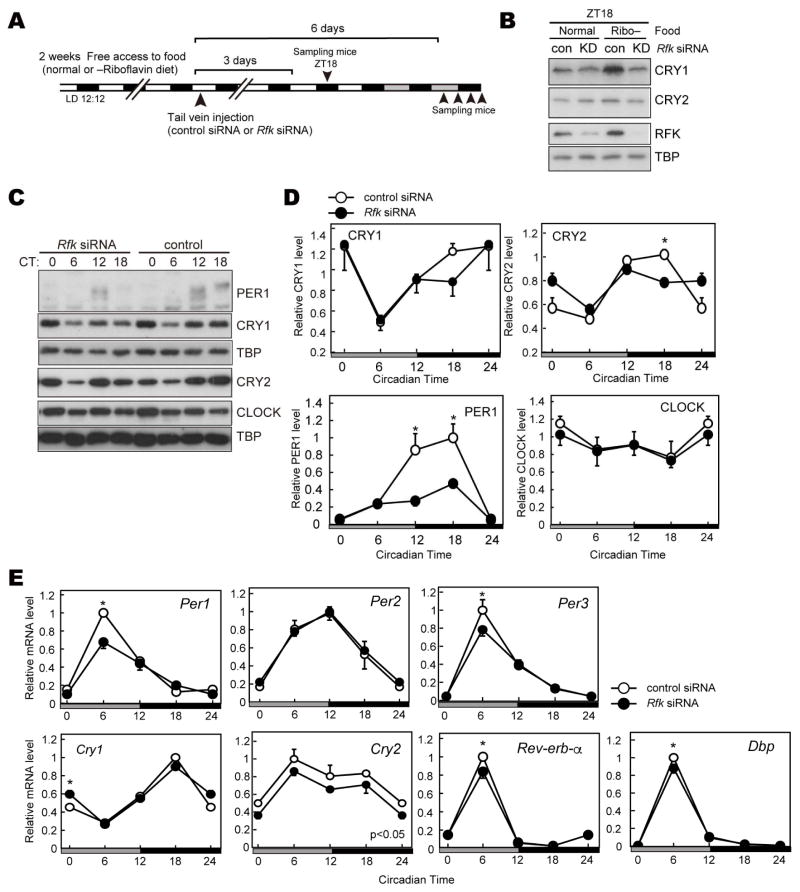
Figure 4. Effect of Rfk knockdown on clock proteins in mouse liver.
(A) Experimental scheme for in vivo knockdown of the Rfk gene. Mice were fed normal or riboflavin-deficient chow for 2 weeks. Rfk siRNA or control siRNA was injected into mice via the tail vein at ZT10-12. Three days or 6 days after the injection, mice were sacrificed to collect liver tissues. (B) Nuclear protein levels of CRY1, CRY2 and RFK at ZT18. TATA-binding protein (TBP) was used as a nuclear marker. (C, D) Nuclear protein levels of PER1, CLOCK, CRY1, CRY2 at indicated CT in DD. Data are shown as means ± SEM (n=3, *:p<0.05 by two-way ANOVA followed by post-hoc test). (E) mRNA levels of indicated clock genes in mouse liver at specified CT in DD. mRNA levels were normalized to G3pdh. Data are shown as means ± SEM (n=3, *:p<0.05 by two-way ANOVA followed by post-hoc test).

