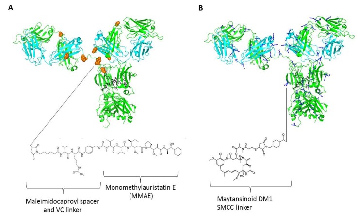
Figure 1.
Structural elucidation of antibody drug conjugates (ADCs) using the IgG crystal structure (PDB code: 1HZH) with two-dimensional drawings of drug molecules with linker and spacers (not drawn to scale). (A) A model of brentuximab vedotin in which conjugation of cysteines via maleimidocaproyl-VC dipeptide-PAB-MMAE is shown. Residues in orange spheres represent the eight naturally occurring cysteines; (B) A model of trastuzumab emtansine in which the drug maytansinoid DM1 is linked to lysines via SMCC thioether. Residues in blue sticks represent the more than eighty naturally occurring lysines.

