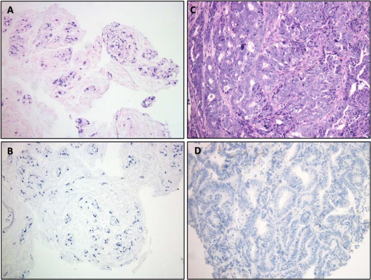
Figure 1.
Histomorphology images from patients 1 and 2. (A): Histologic appearance of hematoxylin and eosin stained tumor from patient #1 demonstrating a discohesive mucinous tumor with signet ring morphology without true glandular formation. (B): No discernable proliferation of tumor‐infiltrating lymphocytes (TILs) was noted, and immunohistochemical staining for programmed death‐ligand 1 (PD‐L1) was negative (DAKO Inc., Carpenteria CA, Clone 22C3). (C): Appearance of tumor from patient 2 reveals a more conventional colonic adenocarcinoma, which was moderately differentiated with cribriform growth and irregular glands. (D): While histologically dissimilar from patient 1, here too, the presence of TILs was insignificant and PD‐L1 staining was negative. Immunohistochemical staining for the mismatch repair proteins was retained in both specimens (not shown). All images taken at ×200 total magnification.

