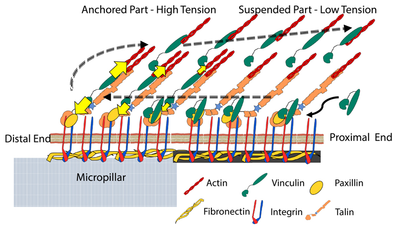
Figure 5.
A model describing the structure of FA on the micropillar top wherein one part of the FA is anchored and a significant portion of the FA is suspended from the pillar. The dashed arrows describe the hypothesized travel of vinculin during FA treadmilling. Intra-molecular tension is higher in the anchored part whereas the protruding part experiences relatively lower tension correlating with the lack of substrate attachment. The shaded fibronectin in the protruding part represents the fibrils formed by the cell (see Figure S4).

