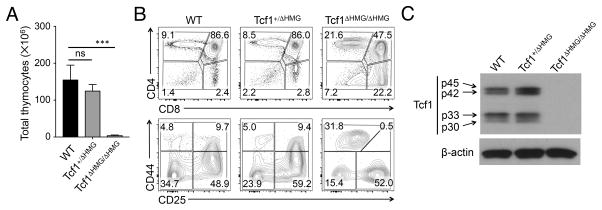
Figure 4. Impact of truncating the HMG domain in Tcf1 on T cell development.
(A) Thymic cellularity in WT, Tcf1+/ΔHMG, and Tcf1ΔHMG/ΔHMG mice (n ≥ 4 from 4 experiments). ns, not statistically significant; ***, p<0.001 by Student’s t test.
(B) Thymic maturation stages. Lin− thymocytes were analyzed for DN, DP, CD4+ and CD8+ populations (top panels in B), and the Lin− DN cells were further analyzed for DN1-DN4 subsets (bottom in B). Contour plots are representative from 4 experiments (n ≥ 4).
(C) Detection of Tcf1 by immunoblotting. Cell lysates were extracted from total thymocytes of control, Tcf1+/ΔHMG, and Tcf1ΔHMG/ΔHMG mice, and immunoblotted with anti-Tcf1 or β-actin antibody. Representative data from ≥ 3 experiments are shown.

