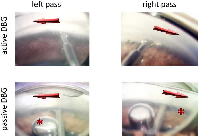
Fig 2. Intraoperative view of active and passive DBG.
The active DBG (top) with an active irrigation and aspiration system. The anterior chamber was stable in both the left and the right pass. A supraphysiological deepening put the trabecular meshwork on tension and in direct view. Debris was aspirated. The passive DBG (bottom) required a viscoelastic to maintain the space. Air bubbles that were trapped in the viscoelastic (left asterisk) could not be removed without retracting the instrument from the eye. The anterior chamber became progressively more shallow as viscoelastic was displaced resulting in a narrowing angle and view obstruction from a billowing iris (right asterisk). Corneal striae from relative hypotony can be seen as well (right asterisk).

