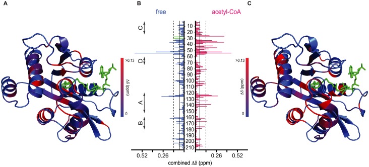
Fig 4. Assembly of bmAANAT3 complexes with acceptor substrates monitored by NMR.
(A) Mapping of tryptophol induced CSPs to free bmAANAT3. The conserved motifs of the GNAT superfamily are indicated on the left. (B) CSPs as a function of primary sequence for the interaction of tryptophol with free- (blue) and acetyl-CoA-bound bmAANAT3 (red). CSPs greater than the mean or one SD above the mean are marked by continuous and broken lines, respectively. Green, open bars in the free-state mark residues that disappear in the substrate-bound state. (C) Mapping of tryptophol induced CSPs to acetyl-CoA bound bmAANAT3. The bisubstrate, CoA-S-acetyltryptamine (green), was modelled by aligning bmAANAT3 model to the structure of oaAANAT.

