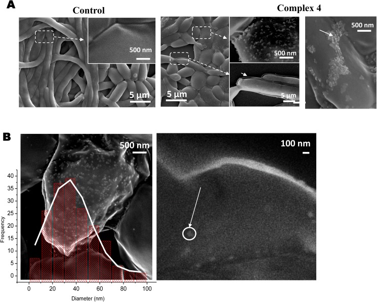
Fig 4. Microscopy images obtained by SEM of C. albicans cells SC5314 when cultivated for 24h in: A1- RPMI growth medium (control); A2- RPMI growth medium supplemented with 125 μg/mL of 4.
Panel A2 shows a magnification of the image evidencing the occurrence of modifications on the cell surface; B- size distribution of the small nanoparticles found at the cell surface of C. albicans.

