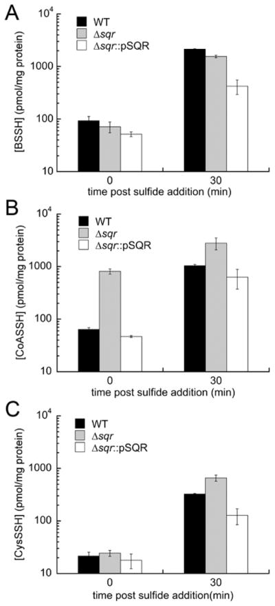
Figure 7.
Cellular LMW persulfide and inorganic sulfide profiling of wild-type (WT), Δsqr, and Δsqr::pSQR S. aureus Newman strains before (t = 0) and after (t = 30 min) addition of 0.2 mM Na2S to early log phase (OD600 = 0.2) cultures: (A) bacillithiol persulfide (BSSH) for WT (black bars), Δsqr (gray bars), and Δsqr::pSQR (white bars) strains, (B) CoASSH, and (C) cysteine persulfide (CysSSH). All cultures were grown in HHWm medium supplemented with 0.5 mM thiosulfate and 10 μg/mL chloramphenicol. WT and Δsqr::pSQR samples were analyzed as biological triplicates, and the Δsqr strain was analyzed as a biological replicate. Analogous data are shown for inorganic hydrodisulfide and tetrasulfide species (Figure S7).

