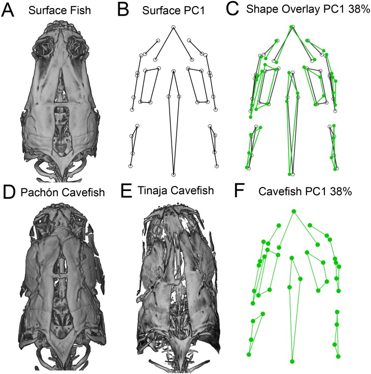
Fig 4. The dorsal osteocranium demonstrates directional asymmetry in adult cavefish.
Surface-rendered microCT images of the dorso-cranium are presented for individual surface fish (A), Pachón (D) and Tinaja (E) cavefish. Landmark-based wireframe graphs for PC1 (capturing 38% of asymmetry shape variation) depict the average dorso-cranial shape for surface fish (B; black lines) and cavefish shape from both populations (F; green lines). Cavefish average shape (green) overlaid on surface fish average shape (black) reveals a dramatic dorso-cranial bend to the left (C). Procrustes ANOVA revealed significant and leftward-biased directional asymmetry in the cavefish dorsocranium (p = 0.0409). The bend in cavefish is most severe in the anterior dorso-cranium inclusive of the premaxilla, supraorbitals and the frontal bone, as well as the dorsal foramen, which extends posteriorly to the supraoccipital bone.

