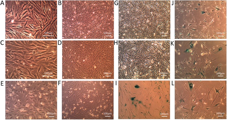
Fig 2. Cell morphology comparisons and β-gal staining of immortalized and primary chicken preadipocytes.
(A) and (B) Light microscopy of ICP1 cells at partial and full confluence at PD 22; (C) and (D) Light microscopy of ICP2 cells at partial and full confluence at PD 15; (E) Light microscopy of the PCPs at partial confluence 1 day after culture; (F) Light microscopy of the PCPs at confluence at PD 2; (G) β-gal staining of ICP1 at PD 100; (H) β-gal staining of ICP2 at PD 100; (I) β-gal staining of the senescent PCPs at PD 9; (J) β-gal staining of chTR retrovirus-infected chicken preadipocytes at PD 10; (K) β-gal staining of empty vector (pLXRN) retrovirus-infected chicken preadipocytes at PD 8; (L) β-gal staining of empty vector (pLPCX) retrovirus-infected chicken preadipocytes at PD 8. Scale bar, 100 μm.

