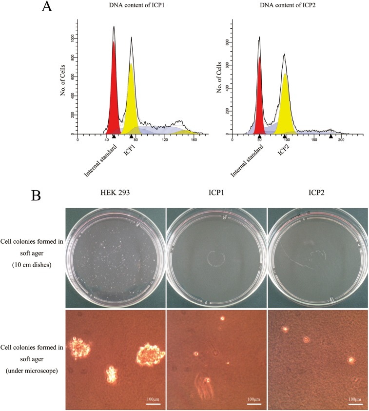
Fig 4. Flow cytometric ploidy analysis and soft agar colony formation assay of the immortalized chicken preadipocytes.
(A) DNA content analysis of ICP1 and ICP2 cells by flow cytometry. PCPs and embryo fibroblasts of AA broiler chickens served as internal standards. (B) Soft agar colony formation assay of ICP1 and ICP2 cells. Cell colonies formed on the dishes were observed by the naked eye, and examined under a microscope. Scale bar, 100 μm.

