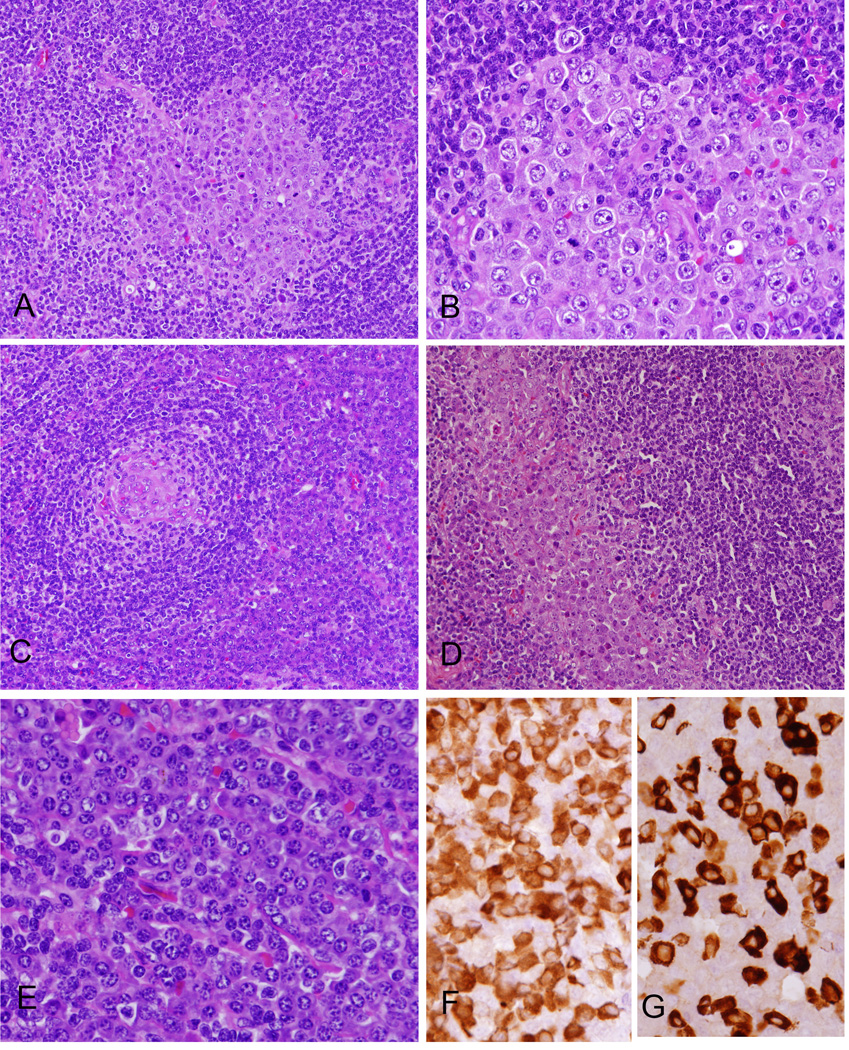
Figure 1. Morphologic features of KSHV- and EBV-associated GLPD.
(A) The atypical cells were largely confined to germinal centers. (B) The cells are large, showing plasmablastic morphology with smooth nuclear contours, vesicular and eccentrically placed nuclei, 1–2 prominent nucleoli and dense amphophilic. (C) Focal Castleman-like areas with regressed germinal centers surrounded by atypical cells are present. (D) Focally the atypical cells infiltrate sinuses, also highlighted with immunohistochemical stains (see Figure 2). (E) There is marked inter-follicular plasmacytosis. Plasma cells are polyclonal for Kappa (F) and lambda (G) light chains by immunohistochemistry, while the plasmablastic cells were negative (not shown).

