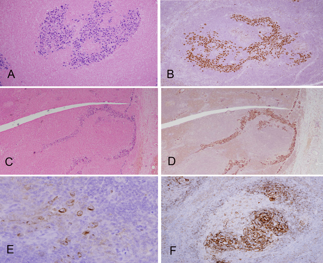
Figure 2. Localization of EBV and HHV-8 in GLPD.
The distribution of tumor cells within colonized follicles is highlighted by EBER in situ hybridization (A) and immunohistochemistry for the latent protein, LANA, of HHV-8 (B). However a sinusoidal distribution was seen focally with EBER (C) and staining for LANA (D). The atypical cells are focally positive for vIL-6 (E). CD23 staining shows an intact FDC meshwork within a partially involved follicle (F).

