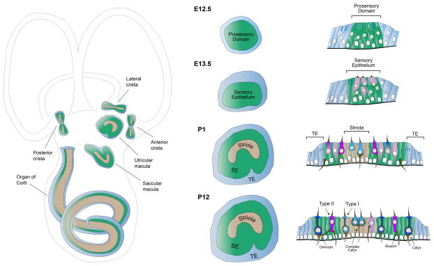
Figure 1. The sensory organs of the mouse inner ear.
The structure of the inner ear sensory organs is shown (left column), as well as the development of the utricular macula in surface (middle column) and cross-sectional (right column) views. The most mature epithelia are shown at the bottom. Left column, Detection of sound or acceleration occurs in the sensory epithelia (green), which are ordered patches comprised of mechanosensitive hair cells and supporting cells. The lateral, posterior, and anterior cristae detect rotational acceleration, the utricle and saccule detect linear acceleration, and the cochlea detects sound. In mammals, each sensory epithelium (green) contains a specialized set of hair cells (tan) that enhance range or sensitivity. In the vestibular organs, these specialized cells are located centrally within the epithelium. Middle and right columns, Surface views and cross-sections depicting development of the mouse utricular macula. By E12.5, a pseudostratified layer of neuroepithelial cells within the otocyst differentiates to form a prosensory domain (green), the precursor to the utricular macula. Neuroepithelial cells surrounding the prosensory domain form the non-sensory transitional epithelium (TE, blue). Prosensory cells exit the cell cycle and begin to differentiate into the first hair cells at E13.5. By birth (P1), progenitors are completing final rounds of cell division. The crescent-shaped striola (tan) has distinguished itself from the surrounding extrastriolar zones (green). Many hair cells display the morphological and electrophysiological characteristics of Type I and II hair cells and have formed connections with vestibular nerve endings. By P12, maturation of the sensory epithelium is nearly complete.

