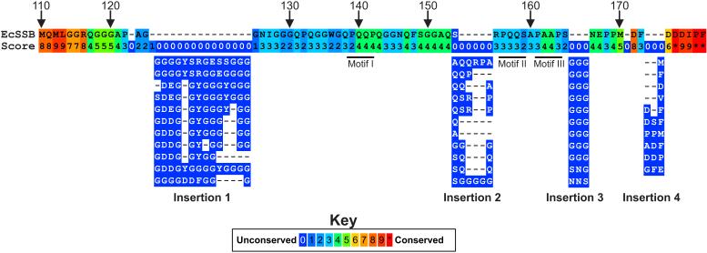
Figure 3. The IDL is not well conserved.
A summary of the alignment of the C-terminal tails of 251 Proteobacterial SSB proteins is presented with only the E. coli sequence shown. The alignment was done using PRALINE (Simossis and Heringa, 2005). The numbering above the sequence corresponds to residue numbering and the values below each amino acid correspond to the alignment score for residues in that position. The positions of the PXXP motifs are indicated by the thick black lines just below positions 140, 156 and 162. Immediately below the E. coli sequence, are representative sequences corresponding to insertions 1-4 (details are in the text). In several cases, the same sequence appeared more than once. Consequently, only one is shown for clarity. The complete alignment is available in Supplementary Figure 1.

