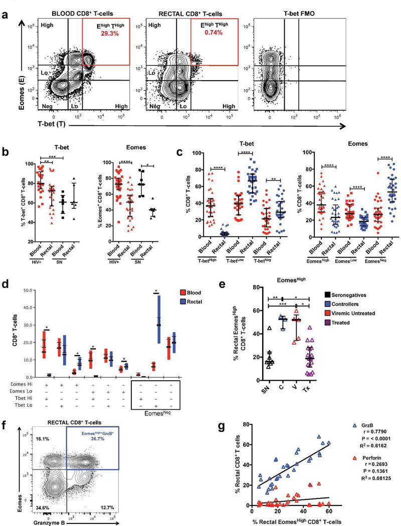
Figure 5.
T-bet and Eomesodermin are differentially expressed in blood and rectal CD8+ T-cells. (a) Representative flow cytometry plot of data from a seronegative participant showing high and low fluorescence intensities of T-bet and Eomesodermin (Eomes) in blood and rectal CD8+ T-cells, highlighting the reduction in frequency of T-betHighEomesHigh CD8+ T-cells in rectal mucosa compared to blood. (b) Differences in the frequencies of total T-bet+ and Eomes+ CD8+ T-cells between blood and rectal mucosa and between HIV+ and seronegative participants. (c) Differences in the frequencies of CD8+ T-cells with T-bet and Eomes high and low expression intensities in blood and rectal mucosa. (d) Difference in T-bet and Eomes co-expression patterns in blood and rectal CD8+ T-cells. (e) Frequency of EomesHigh CD8+ T-cells in rectal mucosa across HIV disease status. Similar results were observed in blood (data not shown). (f) Representative flow cytometry plot showing co-expression of EomesHigh and GrzB in rectal mucosal CD8+ T-cells. (g) Spearman correlation analysis relating the frequency of EomesHigh CD8+ T-cells with the frequency of CD8+ T-cells expressing perforin or GrzB in rectal mucosa. Wide horizontal bars represent medians; narrow whiskers indicate interquartile ranges; asterisks indicate level of significance as follows: * P <0.05, ** P <0.01, *** P <0.001, **** P <0.0001.

