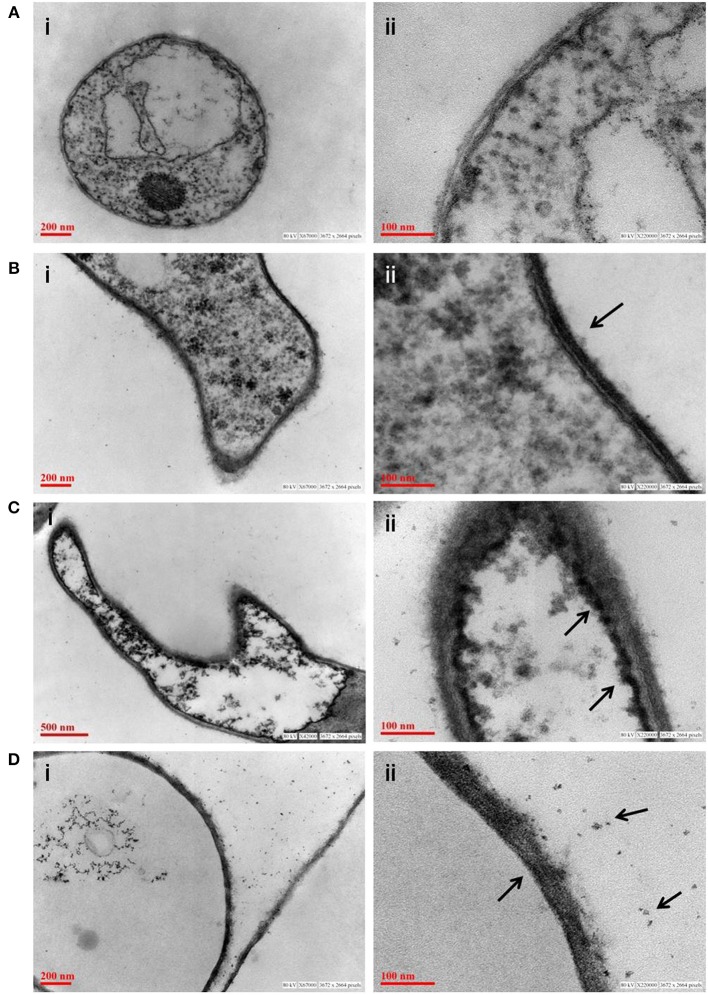
Figure 7.
Transmission electron micrograph of fungus P. indica showing arsenic adsorption, accumulation and precipitation. (A) Untreated fungus; (B) accumulation of arsenic on cell wall of fungus (marked with arrows) treated with sodium arsenate (As V); (C) accumulation of arsenic on in vacuole of fungus (marked with arrows) treated with sodium arsenate (As V); (D) synthesis of insoluble precipitates of arsenic (marked with arrows) on cell wall of fungus treated with sodium arsenite (As III).

