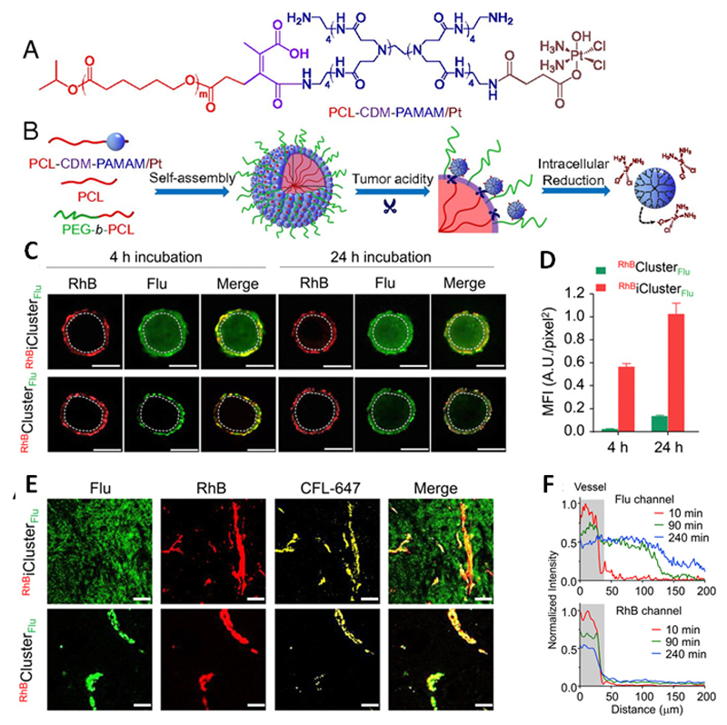
Figure 1.
(A) Chemical structure of PCL-CDM-PAMAM/Pt. (B) Self-assembly and structural change of iCluster in response to tumor acidity. The platinum prodrug-conjugated poly(amidoamine) (PAMAM/Pt) was conjugated to PCL via a pH sensitive linker which was cleaved in the tumor acidic environment, leading to release of the prodrug that showed enhanced tumor penetration due to its smaller size and thereafter intracellular release of Pt. (C) In vitro penetration of RhBiClusterFlu and RhBClusterFlu in cellular spheroids at pH 6.8 after 4- or 24-h of incubation. RhBiClusterFlu and RhBClusterFlu were labeled with fluorescein (Flu, green) on PAMAM and with rhodamine B (RhB, red) on the PCL component. The circles were considered inside area of the spheroids. (Scale bar, 200 μm.) (D) Mean fluorescence intensity (MFI) of green signals (representing PAMAM) in the cellular spheroids. Significantly higher distribution of iCluster inside the spheroids was presented 4-h post-injection compared to that of Cluster. (E) In vivo tumor distribution of RhBiClusterFlu and RhBClusterFlu 4-h post-injection. Flu (green): PAMAM, RhB (red): PCL core and yellow: blood vessels. (Scale bar, 50 μm.) Extravasation of PAMAM in RhBiClusterFlu from tumor vessels to tumor interstitia was observed whereas that of RhBClusterFlu was negligible. (F) Tumor distribution of PAMAM from RhBiClusterFlu. Time-dependent increase of Flu signal (PAMAM) is shown in the upper panel, demonstrating enhanced penetration of PAMAM, and no significant change of the RhB signal was observed (lower panel) pointing to that there was insignificant penetration of the PCL core. Reprinted from reference [30]. Copyright [2016]. United States National Academy of Sciences.

