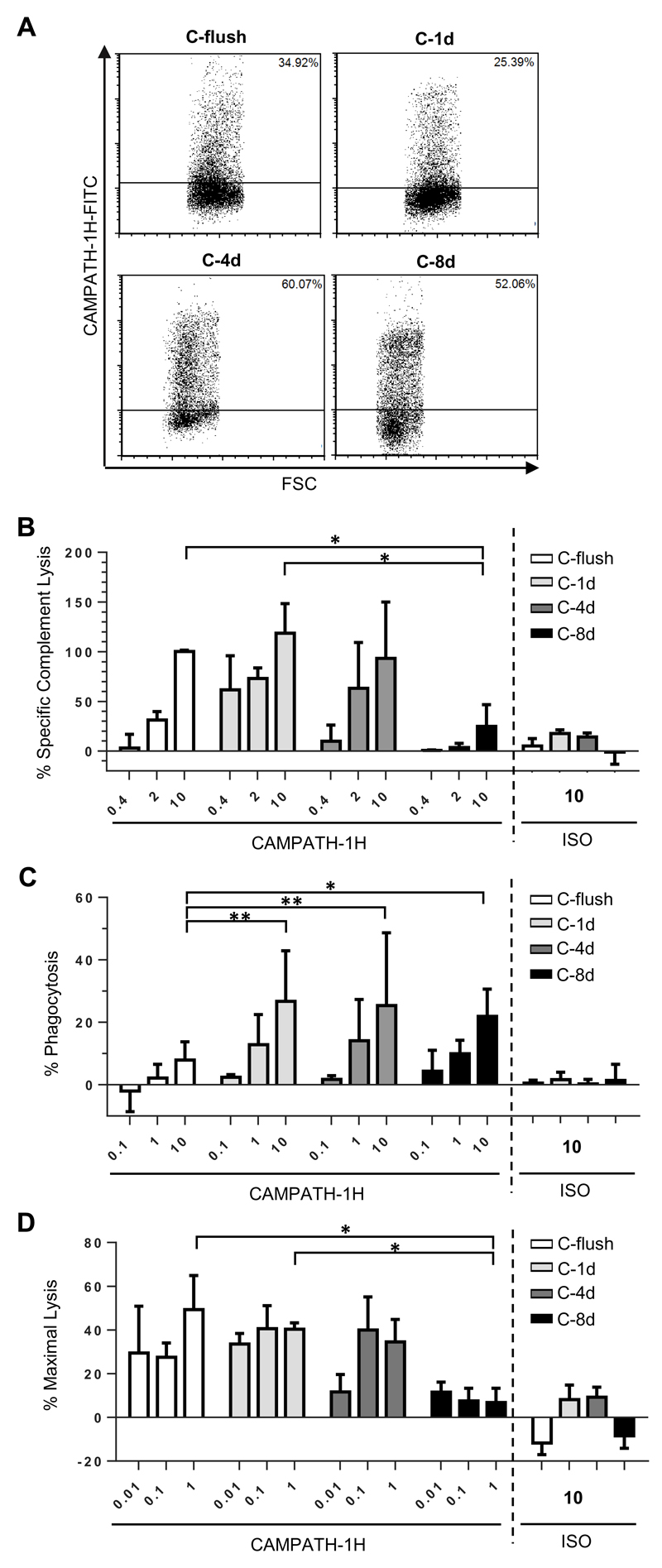
Figure 3. Comparison of cytotoxic activity engaged by CAMPATH-1H when targeting CHO-S cells displaying the CAMPATH epitope at different distances from the plasma membrane.
A) CHO-S cells were transfected with each of the same CD137 backbone constructs as illustrated in Figure 1A but with the rituximab epitope replaced with an epitope recognised by CAMPATH-1H. 24 hours later cell surface expression was confirmed by flow cytometry using FITC-conjugated CAMPATH-1H. CHO-S cells expressing each of the fusion proteins were then used as targets in B) to assess sensitivity to CDC, C) for ADCP and D) for ADCC assay. The mean and SD of three independent experiments are presented. Statistical significance was assessed using a two-way ANOVA test. * p <0.05, ** p <0.005

