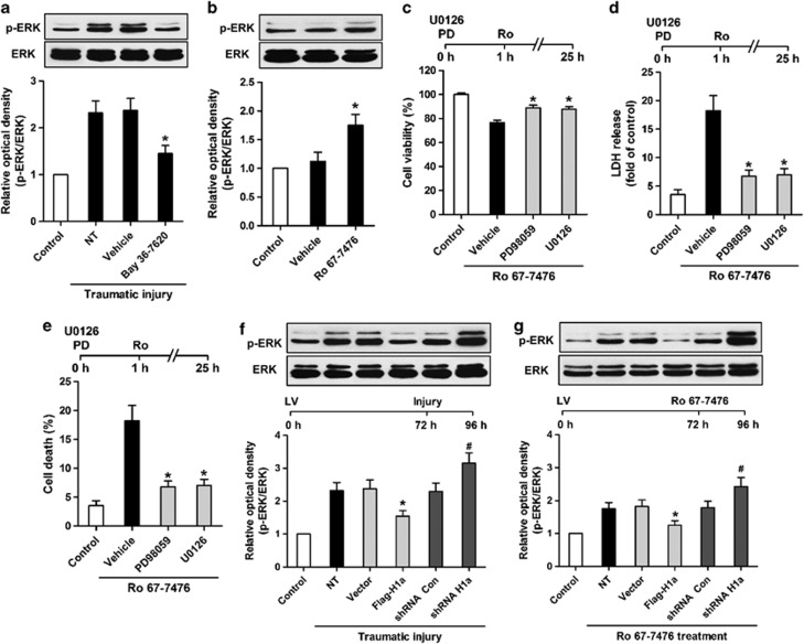
Figure 5.
Homer 1a disrupts mGluR1-extracellular signal-regulated kinase (ERK) signaling. Mice cortical neuronal cultures were pretreated with Bay 36-7620 (10 μM). After traumatic injury, phosphorylation of ERK was measured by western blot analysis (a). The data are represented as the mean±S.E.M. from five experiments. *P<0.05 versus vehicle. Neuronal cultures were treated with Ro 67-7476 (10 μM), and ERK phosphorylation was assessed by western blot analysis (b). The data are represented as the mean±S.E.M. from five experiments. *P<0.05 versus vehicle. Neuronal cultures were pretreated with PD98059 (40 μM) and U0126 (20 μM). Cell viability (c), cytotoxicity (d), and cell death rate (e) were measured in neuronal cultures treated with Ro 67-7476 (10 μM). The data are represented as the mean±S.E.M. from five experiments. *P<0.05 versus vehicle. Mice cortical neuronal cultures were transfected with different lentiviruses, and phosphorylation of ERK was measured by western blot analysis after traumatic injury (f) or treatment with Ro 67-7476 (10 μM) (g). The data are represented as the mean±S.E.M. from five experiments. *P<0.05 versus vector; #P<0.05 versus short hairpin RNA (shRNA) Con

