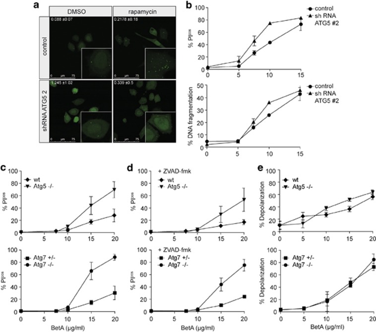
Figure 4.
Autophagy serves as a rescue pathway. (a) HeLa\LC3–GFP cells with pSuper empty (control) or ATG5 #2 knockdown were treated with 10 μM rapamycin for 6 h and analyzed with confocal microscopy. Representative pictures of two experiments are shown. Quantification of LC3 puncta was performed using Image J software. Mean of positive pixels/total pixel cell (%) ±S.D. of three fields of view per sample are shown. (b) HeLa cells with ATG5 #2 knockdown and the control were subjected to different concentrations of BetA for 48 h after which cell death was measured via PI exclusion and DNA fragmentation was assessed. Mean±S.D. of triplicates are shown. (c) Mouse embryonic fibroblasts lacking either ATG5 or ATG7 and their respective control cells were subjected to different concentrations of BetA, and cell death (24 h) using (c) PI exclusion was measured. (d) Mouse embryonic fibroblasts lacking either ATG5 or ATG7 and their respective control cells were subjected to different concentrations of BetA for 24 h in the presence or absence of 20 μM zVAD.fmk, and cell death using PI exclusion was measured. Mean±S.D. of three independent experiments are shown. (e) Loss of mitochondrial activity measured by JC-1 after 18 h was assessed. Mean±S.D. of three independent experiments are shown

