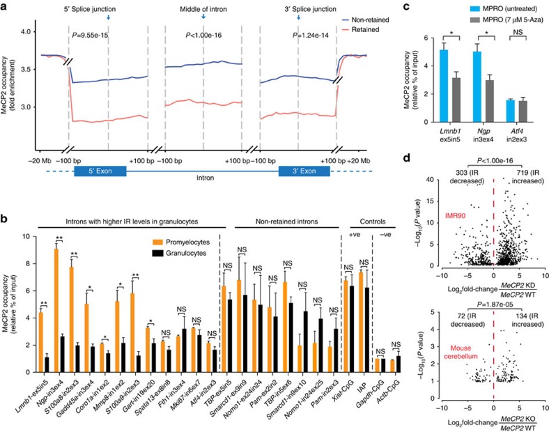
Figure 3. IR is associated with reduced MeCP2 binding near splice junctions.
(a) Average MeCP2 occupancy normalized against input as measured by ChIP-seq in promyelocytes and granulocytes combined. MeCP2 occupancy is displayed for regions spanning ±100 bp from the 5′ and 3′ splice junctions, and the middle of introns, in the DNA encoding non-retained (blue) and retained introns (red). Data were extended to −20 Mb and +20 Mb from the 5′ and 3′ splice junction respectively. t-tests were used to determine significance at P<0.05. (b) Occupancy of MeCP2 near splice junctions of the DNA encoding introns with increased IR in granulocytes and controls measured by ChIP-qPCR. (c) ChIP-qPCR-measured occupancy of MeCP2 near splice junctions of the DNA encoding differentially retained introns of Lmnb1 and Ngp pre- and post- 5-Aza-2′deoxycytidine treatment (5-Aza) in MPRO cells. Atf4 intron 2 is included as a negative control. (d) Association between IR ratio fold changes and their significance following Mecp2 knockdown in IMR90 cells or Mecp2- knockout in primary mouse cerebellum. Binomial test was used to determine the significance of bias between increased and decreased IR at P<0.05. For each ChIP-qPCR experiment, a two-tailed Student's t-test was used to determine significance, denoted by *P<0.05, **P<0.001 and ns, not significant. Bars display mean±s.e.m. Independent ChIP-qPCR experiments were performed three to five times in triplicate.

