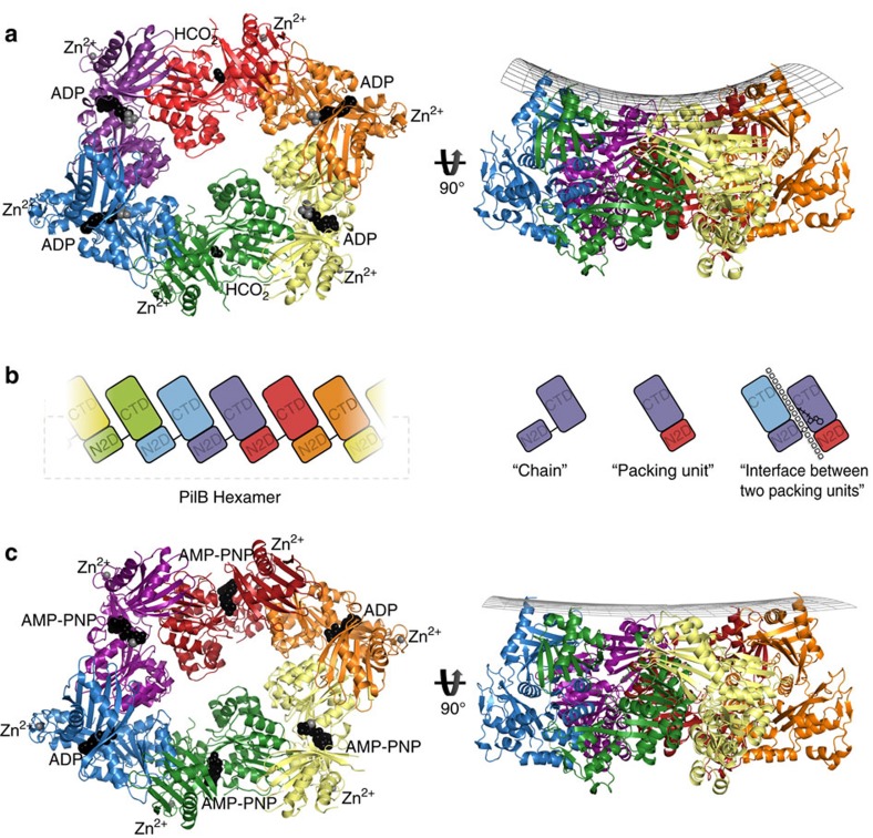
Figure 2. Structure of PilB.
(a) PilB:ADP hexamer, with each chain indicated in a different colour. ADP and formate are shown as black spheres, while magnesium and zinc are shown as grey spheres. A side view is shown with a grid drawn to emphasize the saddle-like shape. (b) Cartoon block illustrations of the PilB hexamer demonstrating the packing units observed, as well as defining the terminology used herein. In the cartoon on the far right, the interface between packing units is represented by a dashed line and the location of the ATP shown in stick representation. (c) PilB:AMP-PNP hexamer, with each chain coloured as in a. AMP-PNP and ADP are shown as black spheres, while magnesium and zinc are shown as grey spheres. A side view is shown with a grid drawn to emphasize the planar shape.

