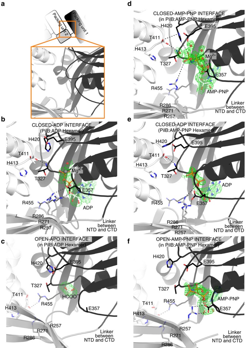
Figure 4. ATP binding sites of PilB.
Direct polar contacts are shown as dashed lines. Magnesium is shown as a grey sphere. The green mesh represents the Feature Enhanced Map computed by PHENIX-FEM45 contoured at 2.0σ. (a) Cartoon clarifying the identity of domains in the following sub-Figures. The cartoon mirrors Fig. 2b. (b) Nucleotide binding site in the closed-ADP interface from PilB:ADP. (c) Nucleotide binding site in the open-APO interface from PilB:ADP. (d) Nucleotide binding site in the closed-AMP-PNP interface from PilB:AMP-PNP. (e) Nucleotide binding site in the closed-ADP interface from PilB:AMP-PNP. (f) Nucleotide binding site in the open-AMP-PNP interface from PilB:AMP-PNP.

