FIG 6 .
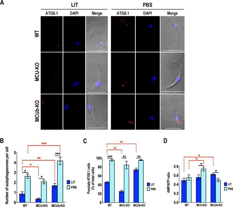
Autophagy changes in mutant epimastigotes. (A) Representative fluorescence microscopy images of wild-type (WT), TcMCU-KO (MCU-KO), and TcMCUb-KO (MCUb-KO) epimastigotes labeled with anti-TcATG8.1 antibody (red) after incubation in LIT medium or PBS for 16 h. DAPI staining (blue) and merged images on a differential interference contrast (DIC) background (Merge) are also shown. Bars, 10 µm. (B) Number of autophagosomes per cell under different conditions shown in panel A. (C) Percentage of cells with autophagosomes under conditions shown in panel A. (D) Comparison of AMP/ATP ratios between WT, MCU-KO, and MCUb-KO epimastigotes incubated in LIT medium or PBS for 16 h. For panels B to D, over 200 cells from wild-type (WT), TcMCU-KO (MCU-KO), and TcMCUb-KO (MCUb-KO) epimastigotes from three experiments with 20 random fields/experiment were analyzed. Values in panels B to D are means ± SD (n = 3). *, P < 0.05; **, P < 0.01; ***, P < 0.001.

