
Figure 2. Vascular and lymphatic vessels in the brain.
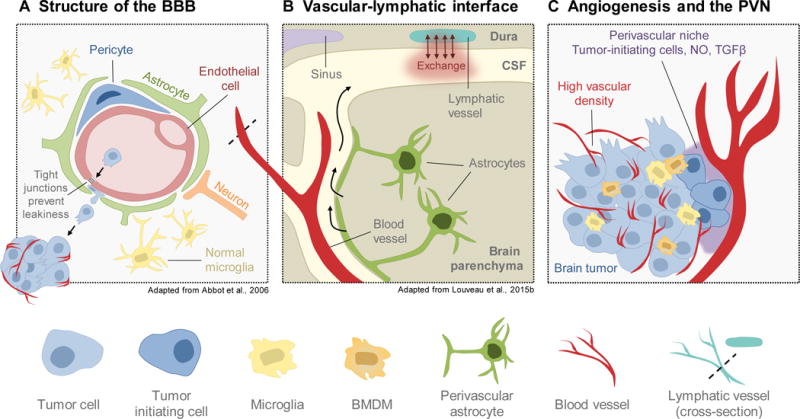
(A) The blood-brain barrier (BBB) serves to protect the brain from inflammation and systemic insults. It is composed of endothelial cells, pericytes, and astrocytes, which tightly seal the endothelium to regulate permeability. Microglia and neurons can additionally contribute to regulation of BBB integrity. Breakdown of junctional integrity can increase permissiveness to seeding of brain-metastatic cancer cells. (B) Recent studies have led to the discovery of lymphatic vasculature in the brain along the dural sinuses in mouse. These vessels exchange fluid with the cerebral spinal fluid (CSF) that surrounds the brain parenchyma, explaining longstanding questions about how immune cells are trafficked into and out of the brain. (C) Brain tumors, particularly gliomas, exhibit extremely high vascularity and angiogenesis. Furthermore, the perivascular niche (PVN) serves as a reservoir for tumor-initiating cells within the brain, which supports tumor outgrowth and aggressive behavior. As such, disrupting the brain vasculature and/or the PVN is of interest for clinical management of brain tumors.