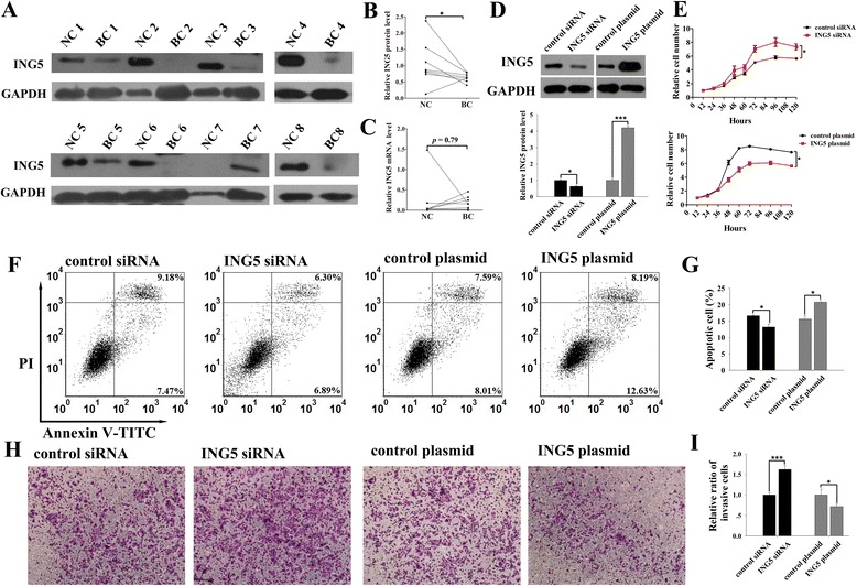
Fig. 1.

Expression levels of ING5 in breast cancer tissues and its role in breast cancer cells. a-b Western blot analysis of the expression levels of ING5 protein in 8 pairs of breast cancer (BC) and noncancerous (NC) tissue samples. a: representative image of the western blot assay; b: quantitative analysis of the western blot assay. c qRT-PCR analysis of the expression levels of ING5 mRNA in 8 pairs BC and NC tissue samples. d Western blot analysis of the expression levels of ING5 protein in MCF-7 cells treated with control siRNA, ING5 siRNA, control plasmid or an ING5 overexpressing plasmid. Upper panel: representative image; lower panel: quantitative analysis. e-i The effects of ING5 on the proliferation, apoptosis and invasion of MCF-7 cells transfected with control siRNA, ING5 siRNA, control plasmid or an ING5 overexpressing plasmid. e: the proliferation curves; f: representative image of apoptosis assays; g: quantitative analysis of apoptosis assays; h: representative image of invasion assays; i: quantitative analysis of invasion assays. *p < 0.05; ***p < 0.001
