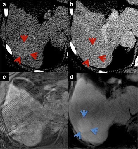
Fig. 6.

Example of patient presenting with early enhancing (a, red arrows) hepatic lesion with rapid contrast wash-out in the portal-venous phase on CT images (b). Additional hepatobiliary phase HSCA-enhanced MRI was acquired to assess lesion uptake. CD-VIBE (c) was non-diagnostic due to motion artifacts in this patient with impaired breath-holding capability. Despite of clearly visible increased lesion uptake on radialVIBE (d, blue arrow), low-grade HCC was confirmed histopathologically following CT-guided biopsy and was subsequently treated by TACE with curative intention
