Fig. 2.

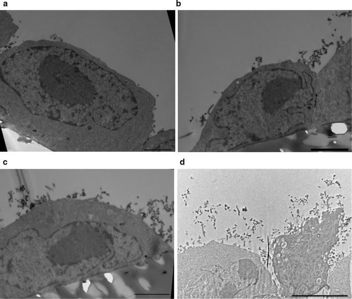
Staining the glycocalyx of CHO WT cells with cationized ferritin shows increasing amounts after several days in culture. Ferritin is seen as spherical dark dots (11 nm in diameter). a One day after seeding CHO WT cells have limited glycocalyx. Most ferritin labels glycans in the intercellular clefts. However, also sparse labelling on the cell surface was noticed. b Similar to a glycans in the intercellular clefts are labelled but after 2 days in culture multiple clusters of ferritin are evident on the cell surface. c After 4 days the cell surface is covered by ferritin staining. d CHO CD36 cells at day 4 after seeding. As for CHO WT cells the glycocalyx covers the entire cell surface. Scale bars show 2 µm in a, b, c and 5 µm in d
