Fig. 3.

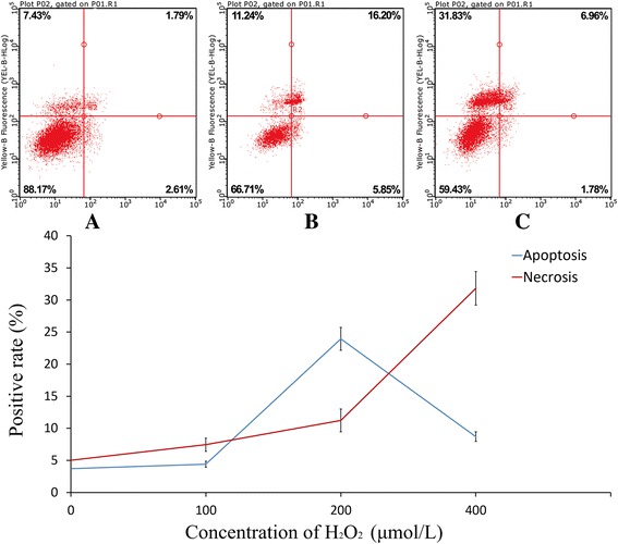
H2O2-induced in vitro cardiomyocyte apoptosis. Cardiomyocytes were treated with 100 μmol/L H2O2 (a), 200 μmol/L H2O2 (b), 400 μmol/L H2O2 (c). Data were expressed as mean ± standard deviation

H2O2-induced in vitro cardiomyocyte apoptosis. Cardiomyocytes were treated with 100 μmol/L H2O2 (a), 200 μmol/L H2O2 (b), 400 μmol/L H2O2 (c). Data were expressed as mean ± standard deviation