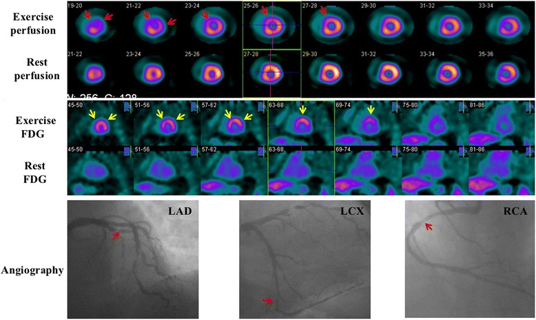
Fig. 4.

Images of a 61-year-old woman. She had a 99% stenosis in the left anterior descending coronary (LAD), a 90% stenosis in the left circumflex coronary (LCX), and a 70% stenosis in the right coronary artery (RCA). Perfusion images show reversible defects in the anterior and lateral wall (red arrows). Resting 18F-FDG images indicate only background uptake in the cardiac cavities and no visible uptake in the left ventricular wall (‘none’ pattern). Nevertheless, exercise 18F-FDG images exhibit intense uptake in the anterior and lateral wall (‘focal’ pattern, yellow arrows) which is in accordance with exercise perfusion images
